Home
Classification
Query
Statistics
Contact
Links
Help
PDB code:
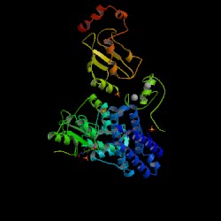
Information on 1fbv
PDB:
1fbv
Compound:
signal transduction protein cbl
Classification:
LIGASE
Entry date in PDB:
2000-08-30
Resolution [Å]:
2.90
R-Factor:
0.227
CHAIN:
A
SWISS-PROT/TREMBL:
P22681
KEYWORD:
3D-structure Calcium-binding Ligase Phosphorylation Proto-oncogene SH2 domain Ubl conjugation pathway Zinc-finger
EC:
6.3.2.-
SCOP:
a.39.1.7
All alpha proteins
EF Hand-like
EF-hand
EF-hand modules in multidomain proteins
Cbl
Human (Homo sapiens)
SCOP:
a.48.1.1
All alpha proteins
N-cbl like
N-terminal domain of cbl (N-cbl)
N-terminal domain of cbl (N-cbl)
N-terminal domain of cbl (N-cbl)
Human (Homo sapiens)
SCOP:
d.93.1.1
Alpha and beta proteins (a+b)
SH2-like
SH2 domain
SH2 domain
Cbl
Human (Homo sapiens)
SCOP:
g.44.1.1
Small proteins
RING/U-box
RING/U-box
RING finger domain, C3HC4
CBL
Human (Homo sapiens)
GO:
nucleus
ubiquitin ligase complex
calcium ion binding
signal transducer activity
ubiquitin-protein ligase activity
zinc ion binding
cell surface receptor linked signal transduction
protein ubiquitination
CHAIN:
C
SWISS-PROT/TREMBL:
P51966
KEYWORD:
3D-structure Ligase Multigene family Ubl conjugation pathway
EC:
6.3.2.19
SCOP:
d.20.1.1
Alpha and beta proteins (a+b)
UBC-like
UBC-like
Ubiquitin conjugating enzyme, UBC
Ubiquitin conjugating enzyme, UBC
Human (Homo sapiens), ubch7
GO:
ubiquitin conjugating enzyme activity
protein modification
ubiquitin cycle
Image Source:
PDB
Homologous structures to 1fbv classified in ArchDB
2cbl
A - percentage of sequence identity: 100